The Basics of Breakthroughs: Approaches in Single-Cell Analysis
We know how important single-cell analysis is to our clients’ research. That’s why as a Contract Research Organization (CRO) specializing in single-cell analysis, we have partnered with top vendors to bring single-cell techniques to our laboratory. Still, researchers may have questions around which platform is best for their study.
This article highlights single-cell technologies, characteristic features, and suitable applications in biological research to help answer your questions. Furthermore, we know that sifting through vendor websites can be time consuming. Contact Canopy Biosciences to speak with a representative about your project or learn more about our CRO services.
What is single-cell analysis?
In 2018, Science named single-cell analysis the “Breakthrough of the Year,” providing some foresight on the popularity it would gain in the coming years. Today, single-cell analysis is one of the most popular approaches in biological research and adoption continues across academic, biotechnology, and pharmaceutical institutions.
While there are a variety of methods and instruments for single-cell analysis, they all have one thing in common; these techniques allow researchers to probe biological processes at the level of individual cells. This level of detail makes possible new biological insights that were previously unattainable using bulk technologies. With single-cell analysis, researchers can study genomics, transcriptomics, and proteomics with a greater level of detail to better understand molecular mechanisms in biological specimens.
Why is single-cell analysis important?
Single-cell analysis is an important tool for researchers who seek to understand molecular mechanisms or gather biological insights at the level of individual cells. Initially, advances in sequencing facilitated the development of single-cell sequencing (scRNAseq), where RNA is extracted from isolated cells to prepare libraries for sequencing.
More recently, image-based approaches have emerged as important tools for single-cell analysis, for their ability to facilitate molecular profiling in situ. In fact, single-cell imaging has proven particularly useful for studying cell-cell interactions within the native tissue microenvironment. The emerging field of spatial biology relies primarily on multiplexed tissue imaging platforms, like ChipCytometry™, to facilitate this type of analysis (1).
What techniques for single-cell analysis does Canopy offer?
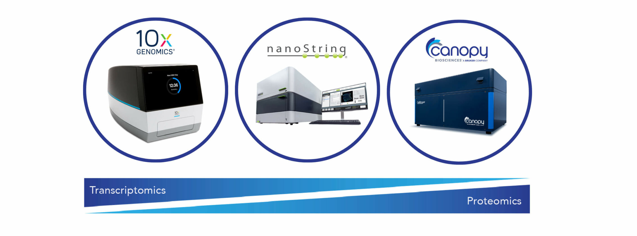
A variety of techniques are available to facilitate single-cell analysis. Canopy Biosciences' CRO arm specializes in single-cell analysis services, including:
- Single-cell RNA Sequencing (scRNAseq) from 10x Genomics: Provides transcriptional profiling for thousands of individual cells to understand gene expression at the single-cell level.
- Spatial Transcriptomics with GeoMx® Digital Spatial Profiler (DSP) from NanoString: Provides spatially resolved information of gene expression according to pre-designed gene expression panels and a limited number of proteins.
- Single-cell Imaging with ChipCytometry™ from Canopy Biosciences: Provides single-cell expression profiles of virtually unlimited proteins.

While scRNAseq and the GeoMx® DSP platform focus on transcriptional profiling, ChipCytometry uses precision imaging to quantify protein levels in tissues and cell suspensions. Each technology offers a different piece of information to help decode molecular mechanisms in health and disease.
Which single-cell analysis technique is best?
Deciding which single-cell analysis technique to use comes down to study design, as each technology offers unique insights. Studies often employ a combination of approaches to compensate for the limitations of any one method. For instance, because cells must be removed from an organism for scRNAseq, it alone can’t show how those cells interact with their neighbors or identify the cells’ descendants.
But by engineering markers into early embryonic cells using CRISPR gene-editing or fluorescent tags – and combining those techniques with scRNAseq – researchers can track cells and their progeny in living organisms. In comparison with scRNAseq, image-based approaches provide transcriptional and proteomic information in intact tissue specimens. Studies that combine transcriptional and proteomic profiling of individual cells can facilitate functional verification of molecular phenotypes.
What is the importance of bulk RNAseq?
Classic bulk RNA sequencing (RNAseq) has been knocked out of the limelight in favor of the more popular scRNAseq. However, bulk RNA sequencing is by no means obsolete. Bulk RNAseq studies provide data on average global gene expression. On the other hand, scRNAseq investigates single-cell RNA biology of up to 20,000 individual cells simultaneously, making bulk RNAseq the method of choice for transcriptomic analysis of pooled cell populations (2).
Because it measures the average expression level across millions of cells for a global idea of gene expression differences, it is ideal for comparing between samples (i.e., healthy vs diseased specimens). Canopy Biosciences offers bulk RNAseq and differential gene expression analysis for studies requiring a more global comparison between samples.
For Research Use Only. Not for use in diagnostic procedures.
References
- Catching up with multiplexed tissue imaging. (2022). Nature Methods, 19(3), 259–259. https://doi.org/10.1038/s41592-022-01428-z
- Li, X., & Wang, C.-Y. (2021). From bulk, single-cell to spatial RNA sequencing. International Journal of Oral Science, 13(1), 36. https://doi.org/10.1038/s41368-021-00146-0