Introducing Enhanced Photobleaching in Cyclic Immunofluorescence for Spatial Biology

Bruker Spatial Biology is pleased to introduce EpicIF™ signal removal technology for the CellScape™ Precise Spatial Proteomics platform. EpicIF empowers high-plex cycling and maximum flexibility in target selection by erasing photostable fluorophores. The gentle method quickly removes fluorescence signals while preserving tissue integrity through an automated process powered by our new CellScape™ Navigator software. EpicIF significantly expands the range of antibody-fluorophore conjugates compatible with multiplex immunofluorescence (IF) experiments and allows scientists to implement a wider variety of fluorescence-based assays, including RNA-ISH and in situ proximity ligation assays, using the CellScape instrument.
What is EpicIF?
Enhanced photobleaching in cyclic immunofluorescence (EpicIF) technology is an expansion of our established filtered photobleaching workflow for multiplex IF signal removal. EpicIF technology enables filtered photobleaching of nearly any organic dye, allowing a wide range of photostable fluorophores from any vendor to be compatible with the CellScape multiplex IF workflow.
EpicIF relies on EpicIF™ Solution, a novel and proprietary reagent that is easily incorporated into the mIF workflow via the automated fluidics system of the CellScape instrument. EpicIF Solution enables the gentle removal of fluorescence signal from fluorophores without damaging tissues or epitopes. Additionally, the reagent is non-toxic, non-hazardous, and easily incorporated into a chemical inventory.

Advantages of EpicIF
EpicIF confers a number of benefits over other multiplexing technologies.
Universality: Use almost any organic dye on the CellScape platform: EpicIF can erase the fluorescent signal from dyes in the Rhodamine, Cyanine, and BODIPY families. Contact us for more information about compatible dyes.
Flexibility: The ability to erase signal from nearly any organic dye, without having to alter the chemistry of the detection probes or the experimental sample, enables the CellScape platform to combine multiplex readouts from different modalities on a single sample. The CellScape instrument can now pair in situ proximity ligation technology (isPLA) and RNA-ISH technology with Precise Spatial Proteomics. Check out our poster highlighting the use of isPLA with CellScape.
Ease of High Plex Assay Design: With detection of up to five fluorophores in each round of staining and imaging and no known limits to the number of cycles possible, the CellScape platform enables truly high plex spatial biology research. The universality of fluorophore compatibility also enables the use of antibodies already validated in other workflows, eliminating the need for additional antibody conjugations and saving time on assay design and optimization.
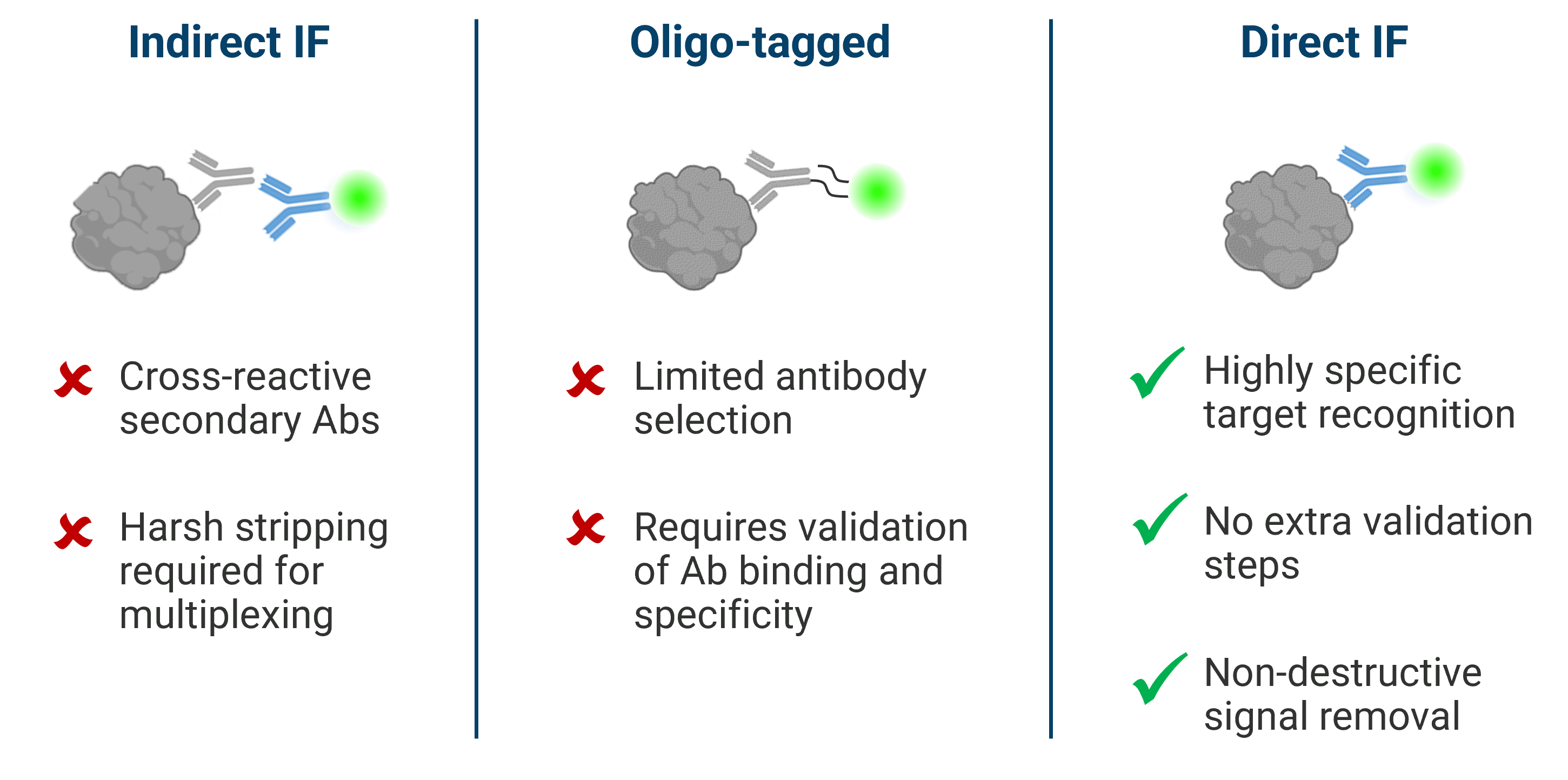
Reliability: The use of primary antibody-fluorophore conjugates facilitated by EpicIF avoids the non-specific background signal and harsh signal removal conditions when using detection methods that rely on secondary antibodies. The non-destructive nature of the EpicIF process preserves epitope stability and tissue integrity, ensuring staining results that are target-specific and trustworthy.

EpicIF Enables High Multiplex
Achieving highly multiplexed immunofluorescence has never been easier.
- Using primary conjugated antibodies with nearly any organic dye and comprehensive signal removal, antibody selection and assay design is simple.
- Built-in High Dynamic Range (HDR) imaging enables automatic exposure optimization for streamlined assay design.
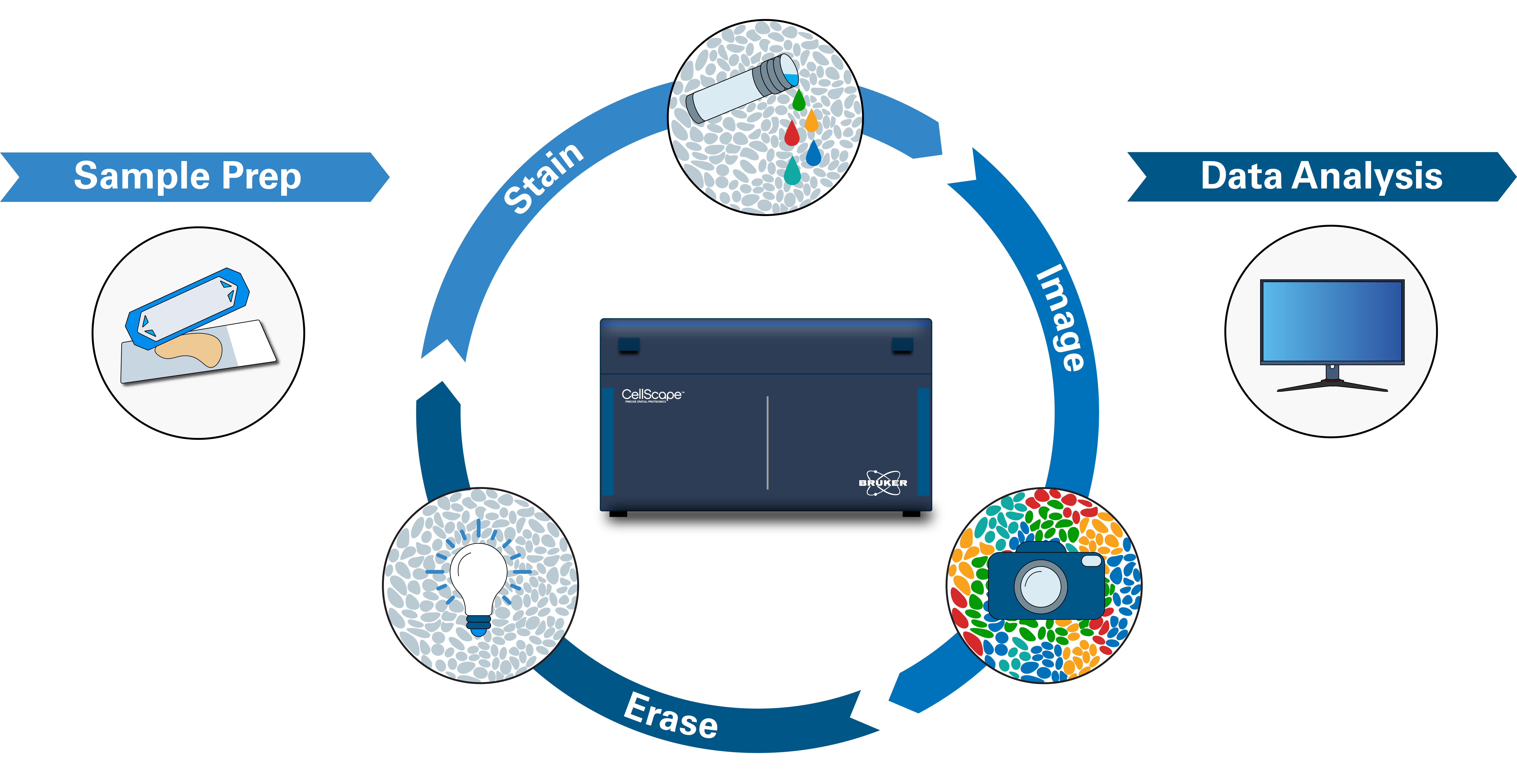
- The CellScape instrument, with its integrated fluidics unit that can hold up to 75 antibodies at a time, controls all liquid handling, staining, imaging, and signal erasure with walkaway automation.

EpicIF is Gentle and Non-damaging
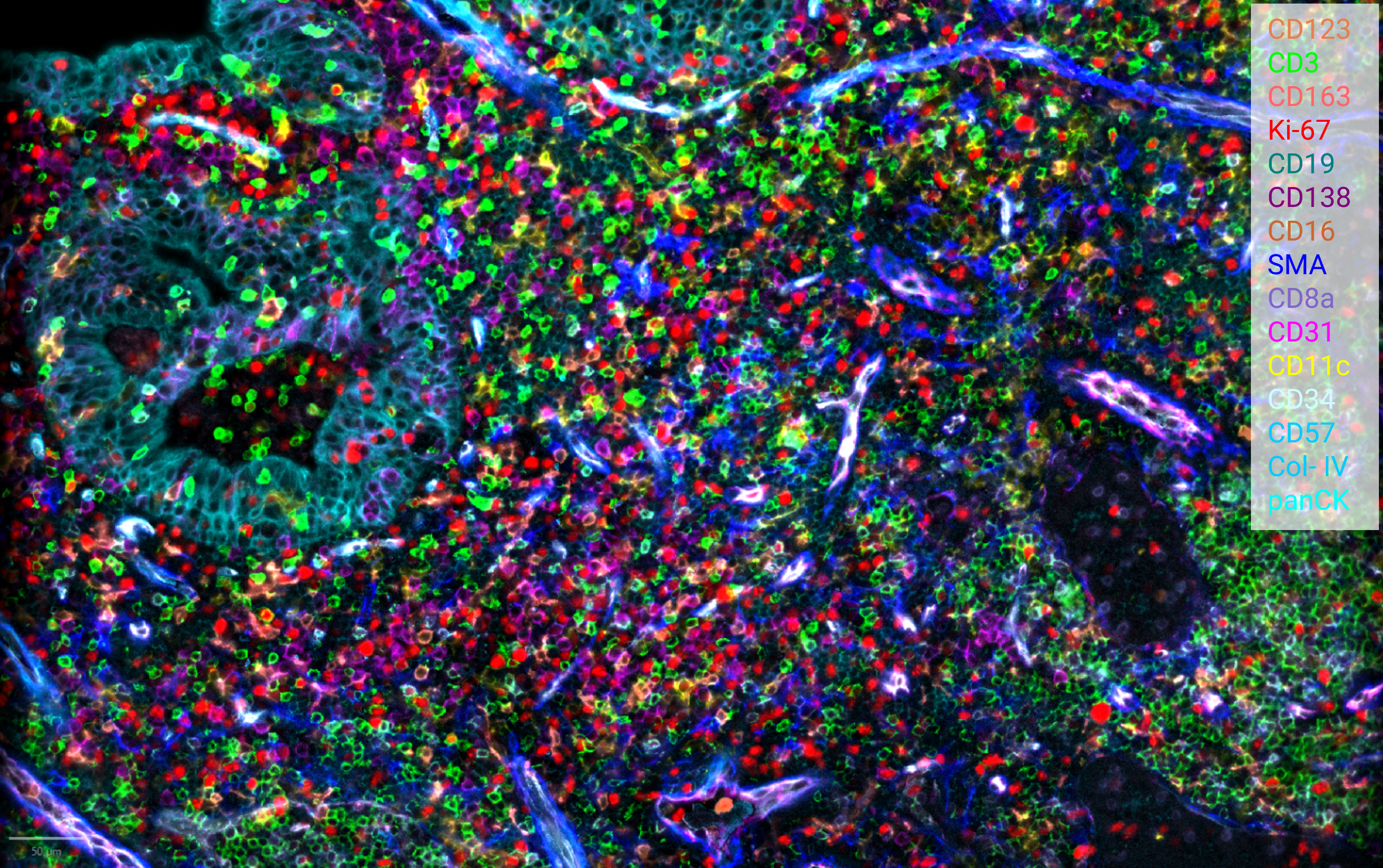
Use of the EpicIF workflow is non-destructive to epitopes, as illustrated in the above image where notoriously difficult-to-detect markers, including FoxP3 and CD279, exhibited clear staining even when included in later cycles.
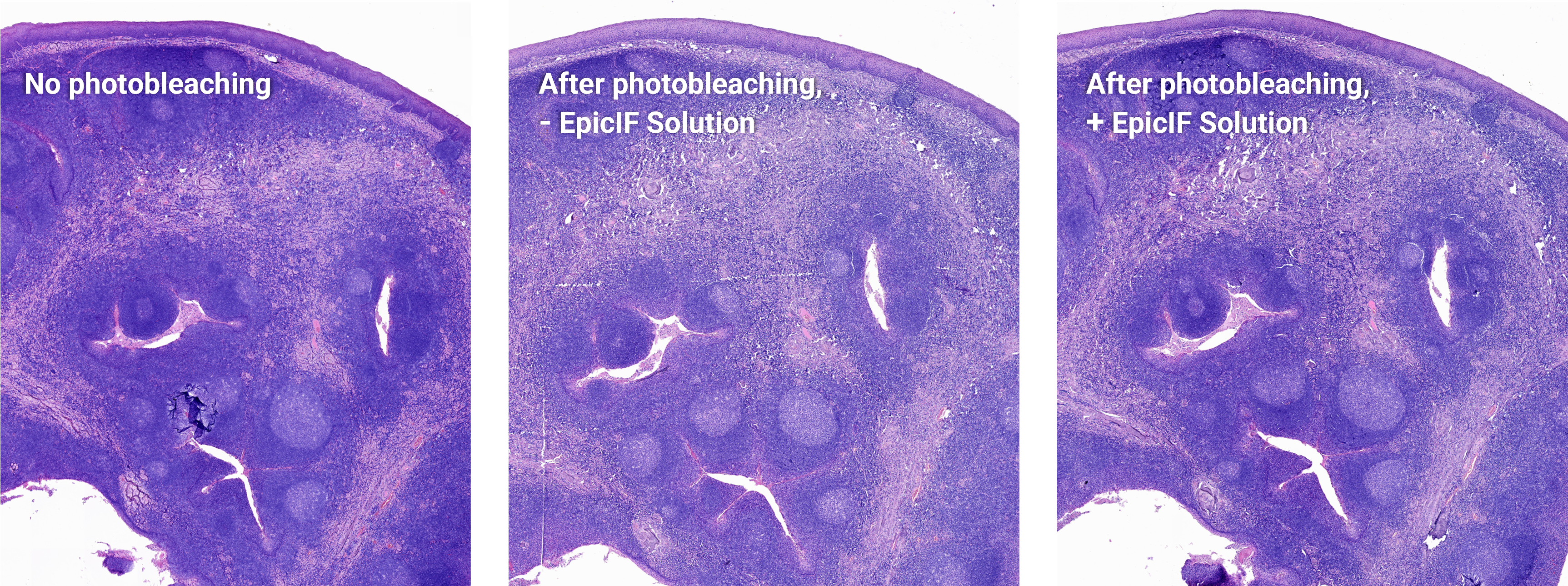
Tissue integrity also remains intact, as illustrated by H&E staining after 10 rounds of EpicIF-mediated photobleaching.

EpicIF is Effective
In most cases, removal of fluorescence signals using EpicIF is complete within 15 seconds, with little to no fluorescence signal remaining, regardless of fluorophore. This exhaustive signal removal allows high multiplexing with minimal signal cross-reactivity, enabling accurate and quantitative cell phenotyping.

Time-lapses of fluorescence signal during 12 seconds of filtered photobleaching with EpicIF solution illustrates complete signal removal. Shown first is human tonsil tissue stained with anti-CD20-AF647 and exposed to light, with and without EpicIF solution to rapidly remove fluorescent signal. Next is the same tissue stained with Sytox Orange (for DNA) and exposed to light, with and without EpicIF solution.
How to Access EpicIF Technology
The EpicIF capability can be accessed starting in Q1, 2025 through upgrades to existing CellScape systems, new instrument purchases, or through the spatial biology CRO capabilities of Canopy Multiomic Services.
If you have additional questions about EpicIF technology, please contact us.
Send us a message
Fill out this form and a member of our team will contact you with more information ASAP.