CellScape™ Precise Spatial Proteomics
High Resolution Quantitative Phenotyping
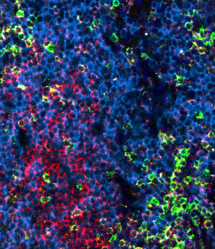
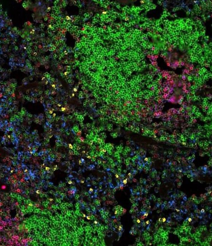
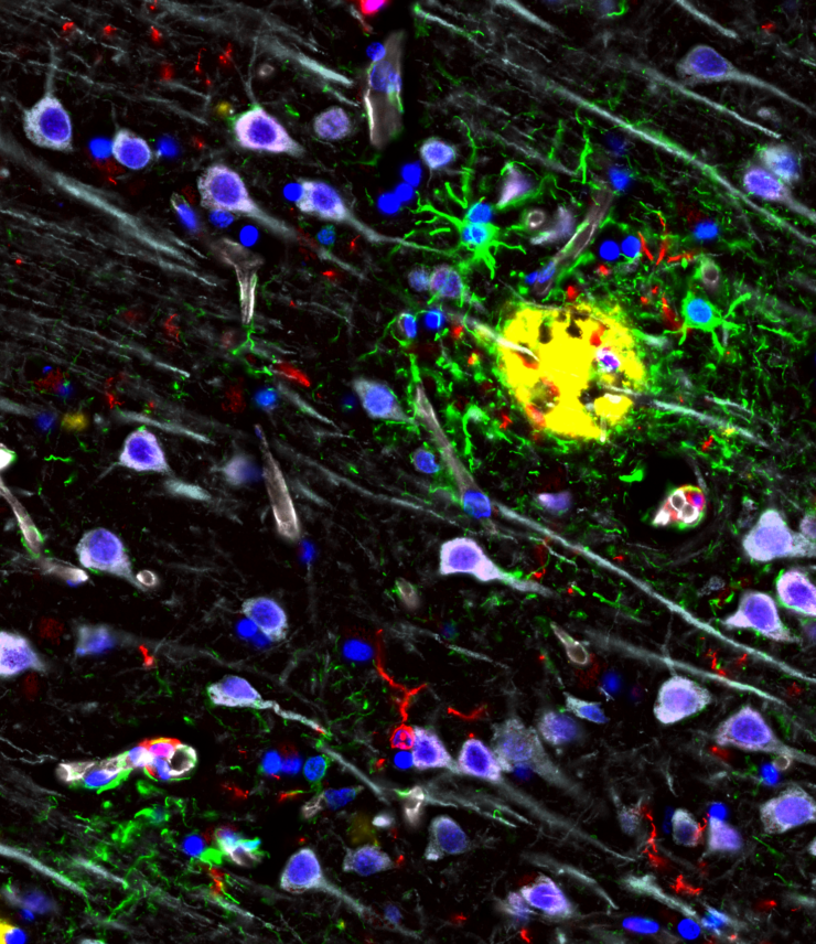
Bruker Spatial Biology brings you best-in-class resolution paired with high dynamic range imaging for robust, high-plex spatial biology. With CellScape, see the big picture, the small details, and everything in between.

NEW: Enhanced Photobleaching in Cyclic Immunofluorescence (EpicIF)
Gentle yet powerful signal removal, compatible with nearly any fluorophore to enhance your unique high-plex spatial proteomics research.
Explore CellScape
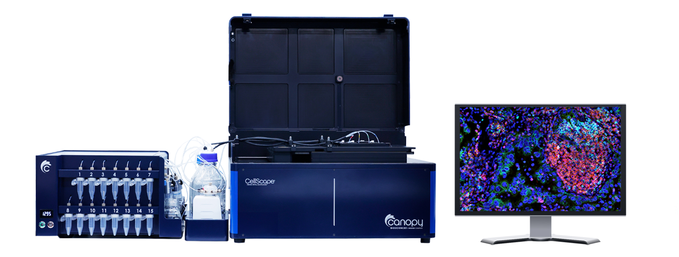
A 4-sample holder and imaging areas of up to 710 mm2 on standard microscope slides enable high-throughput spatial biology.
With a standard image file output (OME-TIFF), data are compatible with both commercially available software and custom image analysis pipelines.
With a digital sampling rate of 182 nm/pixel and high dynamic range (HDR) microscopy, advanced optics provide exceptional data quality.
Integrated microfluidics unit automates immunostaining and ensures reliable reagent delivery.
Fifteen reservoirs for antibody cocktails enable high multiplexing and flexibility in experimental design.
How our Technology Works
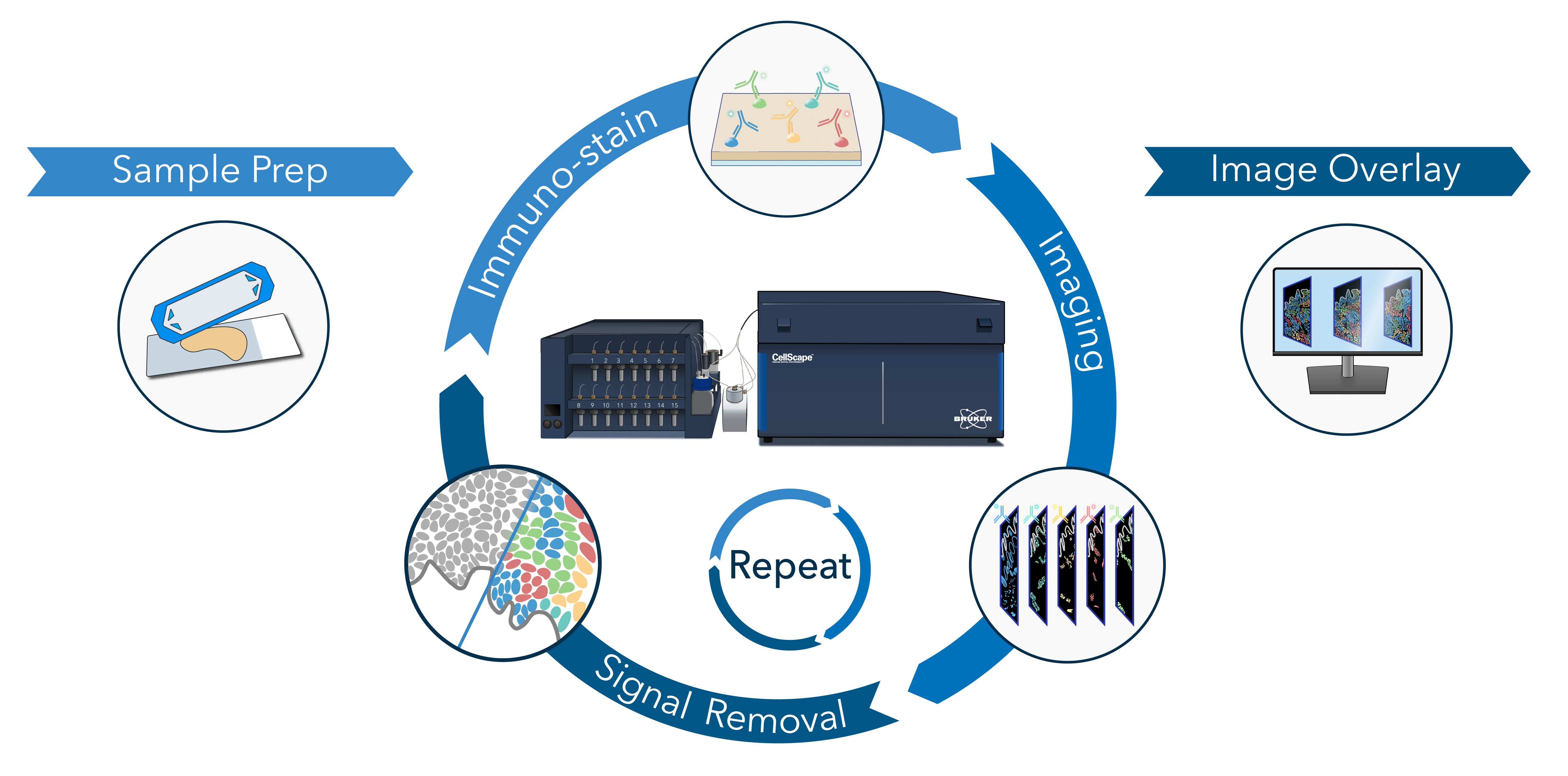
Use our CellScape™ Whole-Slide Imaging Chamber with standard microscope slides to easily prepare tissue and cell suspensions samples from both humans and model organisms.
Simultaneously immuno-stain your sample with up to 5 fluorescent conjugated primary antibodies using off-the-shelf antibodies, our database of validated markers, or our VistaPlex™ Multiplex Assay Kits.
Industry leading optics combined with HDR microscopy achieves true single cell resolution and biomarker quantification of both low and high expressing markers in the same sample.
Photobleaching is used to gently eliminate fluorescence signals, facilitating additional cycles of immunostaining and imaging while ensuring tissue integrity is preserved.
A multiplex image overlay is created by aligning each channel to a reference, enabling simultaneous localization and visualization of multiple biomarkers.
Utilize unlimited cycles to achieve highly multiplexed biomarker detection on your sample.
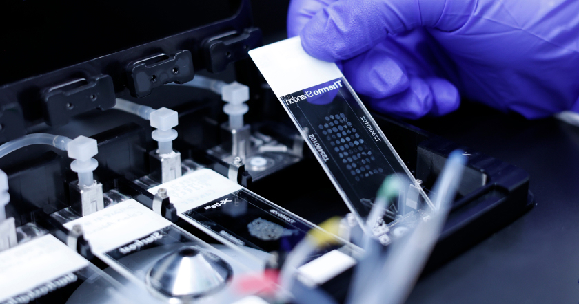
Introducing CellScape Whole-Slide Imaging Chamber
Convert any standard microscope slide into a microfluidic chamber with 710 mm2 available imaging area.
- Increase throughput with more samples on the same slide
- Assay multiple tissue types simultaneously
- Seal, store, and re-explore for modular assay expansion


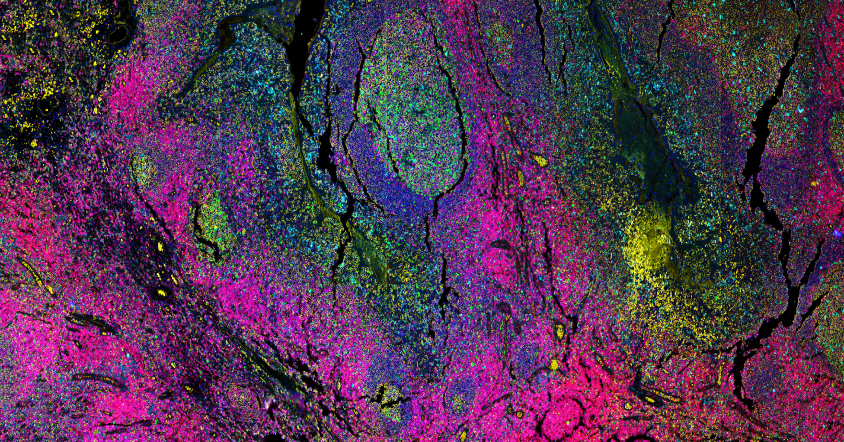
See More Signal
View the entire landscape with High Dynamic Range (HDR) Microscopy
CellScape utilizes an automatic image acquisition process that acquires a series of images across a range of relevant exposure times. As a result, a broader dynamic range of signal is detectable compared to other spatial biology systems that only capture a single exposure.
Additionally, an optimal exposure for each individual biomarker is collected during an experiment, overcoming the need for manual optimization of exposure times for each marker.
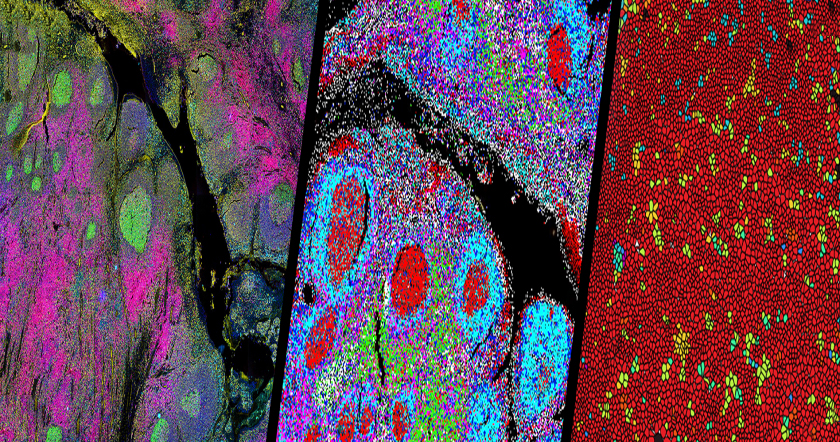
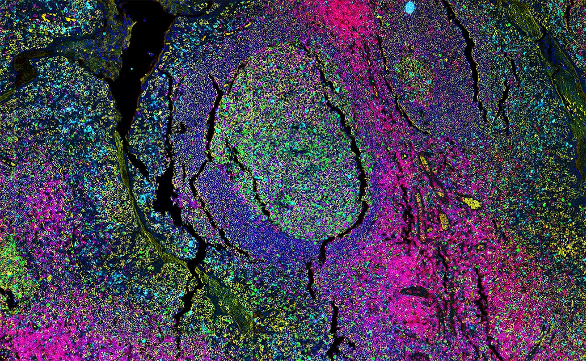
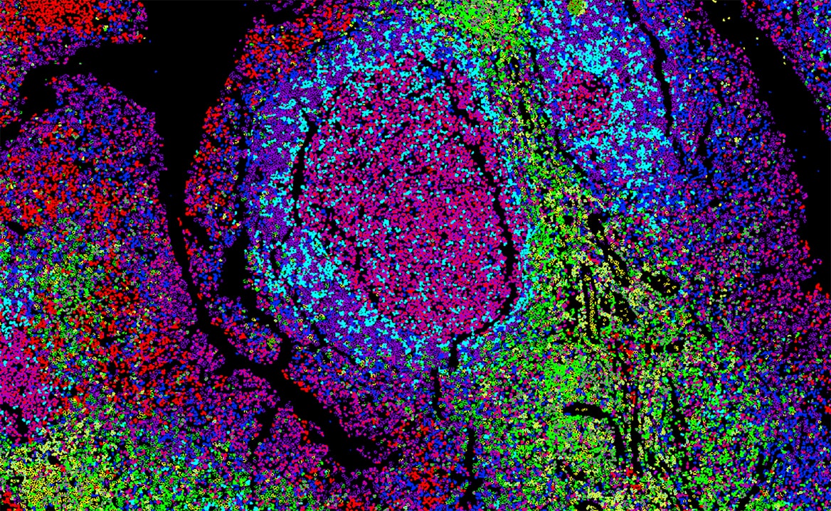
See More Markers
CellScape generates high-plex spatial biology datasets, facilitating unbiased single-cell phenotyping in situ and accelerating biological discovery, therapeutic development, and precision medicine. Use the slider to view a multiplex image generated by CellScape (Left) and the same image after single-cell phenotyping (Right).
See the Advantages for Yourself
View whole-tissue images and insights collected with CellScape Precise Spatial Proteomics using our interactive Data Explorer, powered by Minerva.
Browse through two different highly multiplexed cancer datasets.

See More with Higher Throughput
With two imaging modes to choose from, CellScape allows you to match your throughput needs while maintaining single cell resolution.
Flexible Assay Chemistry
Open-ended assay design allows you to use commercially available fluorescent antibody to build your assay from the ground up and reach your desired multiplex level.

Validated Kits
Ready-to-use, expandable VistaPlex Multiplex Assay Kits give you a jump start on assay design.


Use Your Antibodies
Compatible with fluorescently labeled antibodies from any vendor.
Request a Quote
Interested in purchasing a CellScape for your research?
CellScape Spatial Services
Save time and gain insights by leveraging our in-house expertise. Design your panel with help from our experts, submit your samples, and leave the rest to us.

Related Product

VistaPlex Assay Kits
Leverage VistaPlex Assay kits with validated, ready-to-use antibodies and optimized protocols to accelerate the development of your multiplexed assays.
Featured Publications
Still Have Questions?
Reach out to your local Bruker Spatial Biology Field Application Scientist, contact us or email support.spatial@bruker.com for assistance.