CosMx® Human Whole Transcriptome Pancreas Dataset
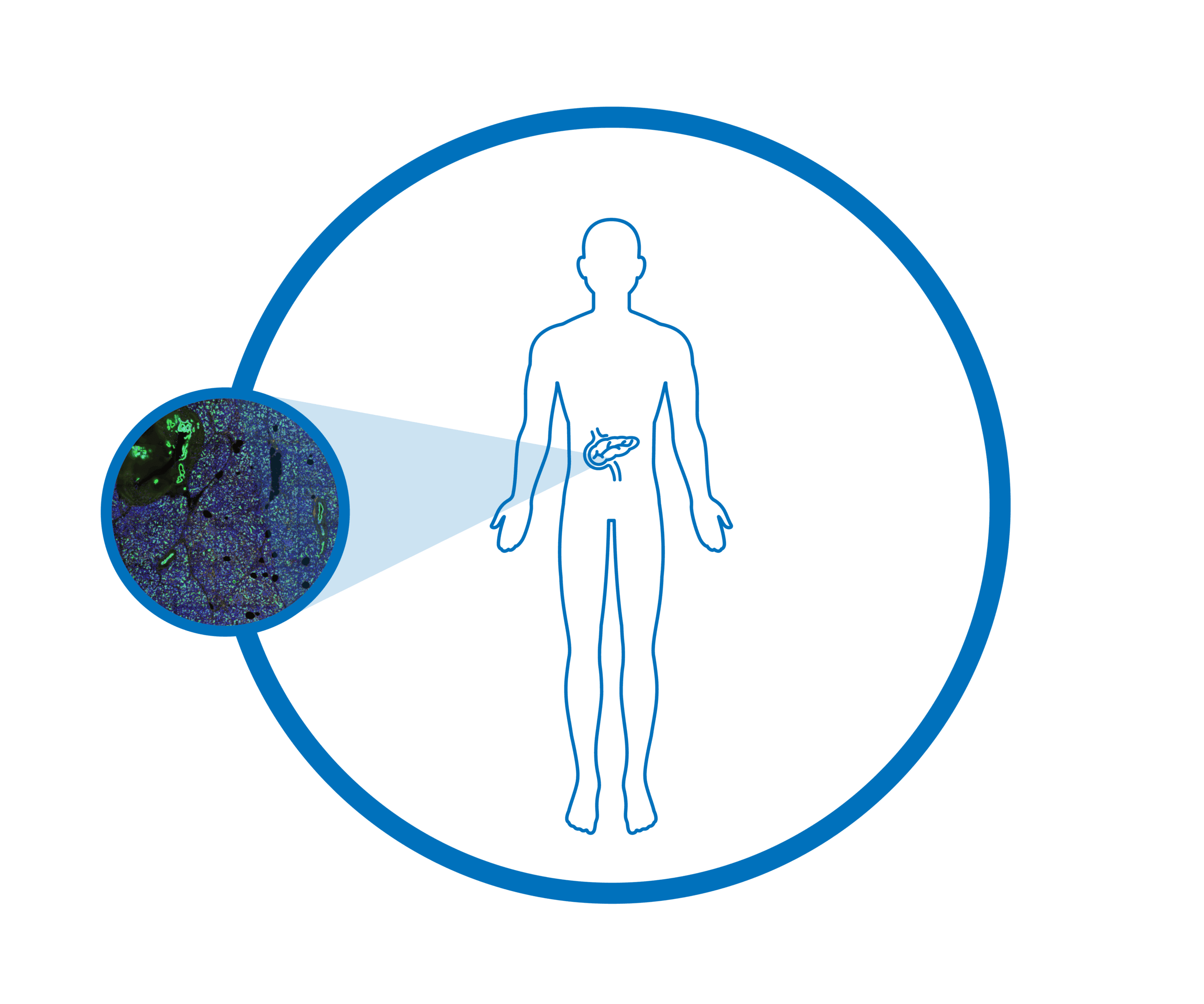
This dataset demonstrates the power of the CosMx SMI in characterizing the biological landscape of the human pancreas. Utilizing CosMx Human Whole Transcriptome Panel, we analyzed normal pancreas tissue from a 55 year old Caucasian female donor. By applying this high plex assay to FFPE tissue, we captured the entire protein encoding transcriptome at the subcellular resolution. This level of detail provides the most comprehensive biological story of pancreatic tissue architecture in a donor with a complex clinical history including hypertension and pulmonary fibrosis. By visualizing the distribution of the whole transcriptome in these samples, researchers can identify unique molecular signatures and cellular interactions within healthy tissue, paving the way for a deeper understanding of human physiology and disease development.
The CosMx® SMI and decoder probes are not offered and/or delivered to the Federal Republic of Germany for use in the Federal Republic of Germany for the detection of cellular RNA, messenger RNA, microRNA, ribosomal RNA and any combinations thereof in a method used in fluorescence in situ hybridization for detecting a plurality of analytes in a sample without the consent of the President and Fellows of Harvard College (Harvard Corporation) as owner of the German part of EP 2 794 928 B1. The use for the detection of cellular RNA, messenger RNA, microRNA, ribosomal RNA and any combinations thereof is prohibited without the consent of the President and Fellows of Harvard College (Harvard Corporation).
*defined-by: (transcripts per plex per cell) / (negatives per plex per cell)
Download Data
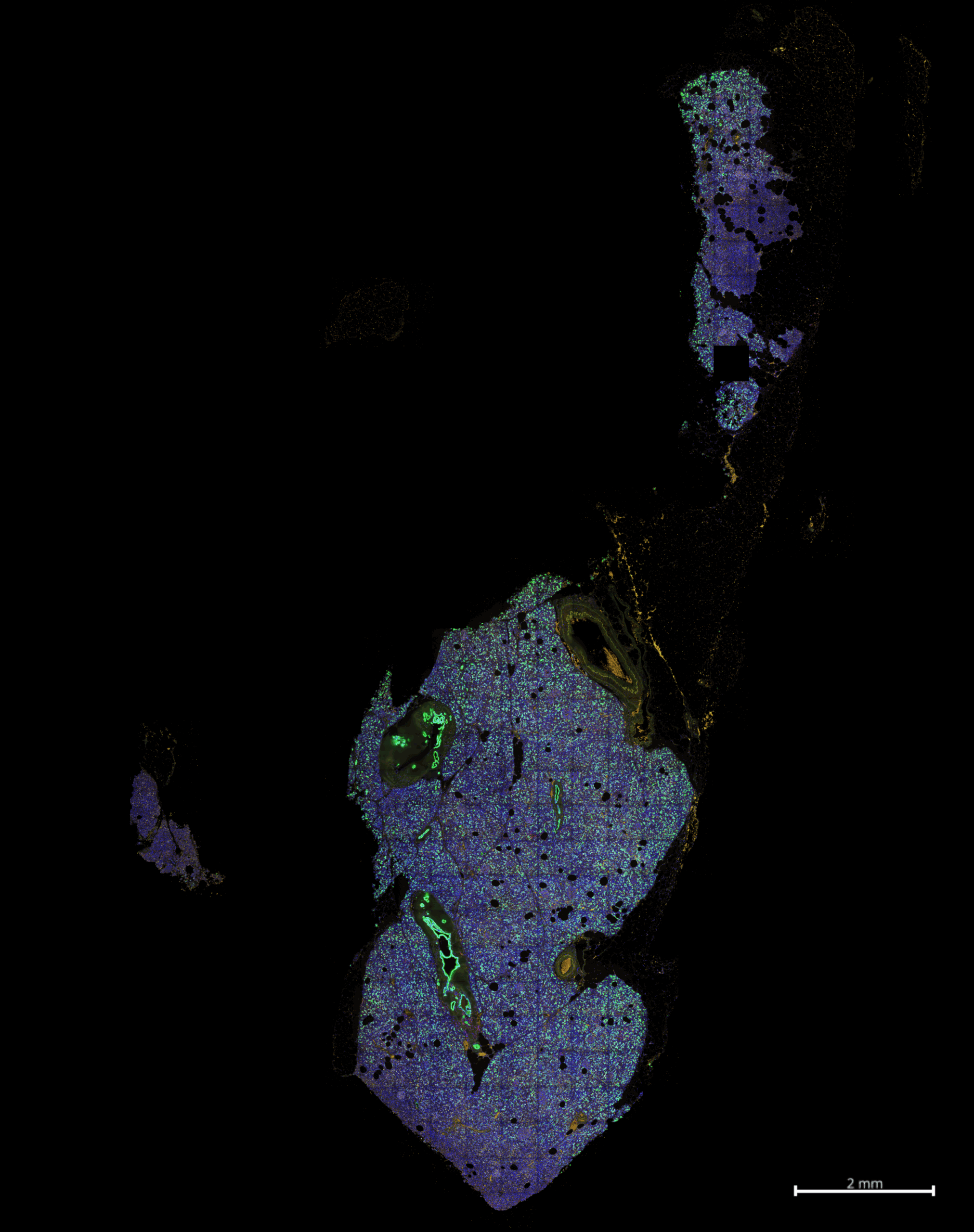
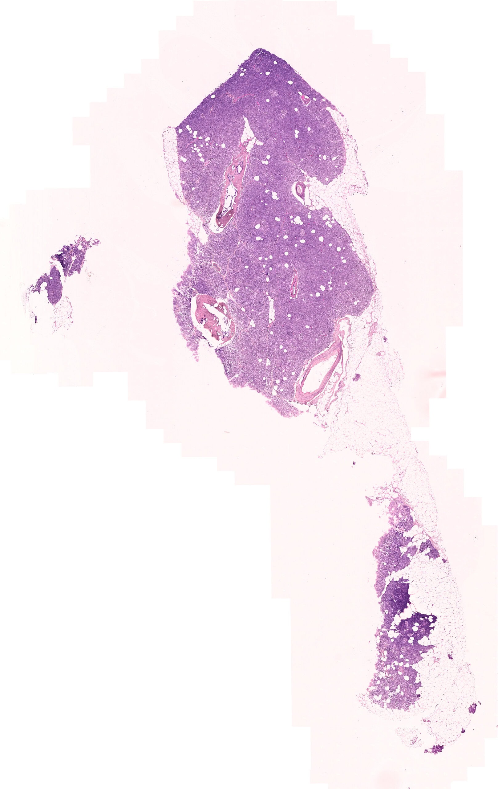
Dataset Overview
Whole transcriptome spatial data from pancreas tissue. The immunofluorescence image below shows CD45 in red, CD68 in yellow, DNA in blue, membrane in gray, and PanCK in green. An H&E image is also available for comparison.
Request More Information
Contact our helpful experts and we’ll be in touch soon.

Technology Publication:
High-Plex Multiomic Analysis in FFPE Tissue at Single-Cellular and Subcellular Resolution by Spatial Molecular Imaging