


CosMx Spatial Molecular Imager
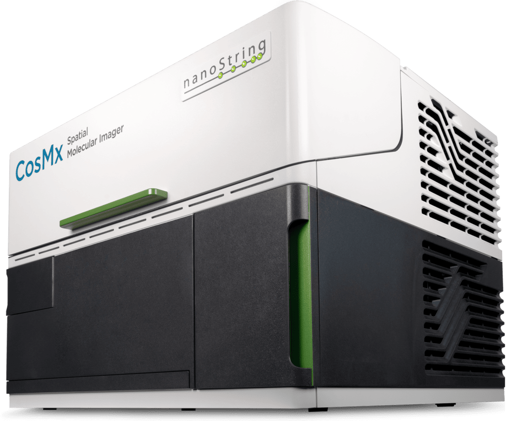
Study whole transcriptome biology at the subcellular level with spatial molecular imaging
CosMx® Spatial Molecular Imager (SMI) is the first high-plex in situ analysis platform to provide spatial multiomics with formalin-fixed paraffin-embedded (FFPE) and fresh frozen (FF) tissue samples at single cell and subcellular resolution.
Single-Cell Imaging at Subcellular Resolution
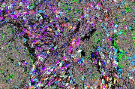
CosMx SMI delivers whole-transcriptome spatial imaging and same-cell multiomics with unmatched sensitivity at single-cell and subcellular resolution.
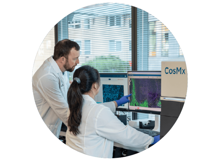
Single Cell Spatial Transcriptomics 101: Intro to CosMx SMI
Understand why spatial biology matters, explore the value of 1K, 6K, and WTX CosMx SMI assays, and determine the right assay for your study.
Explore a Human Whole Transcriptome Breast Cancer Atlas
Explore CosMx SMI enabled human whole-transcriptome spatial biology in breast cancer at single-cell and subcellular resolution.
What sets CosMx SMI apart?
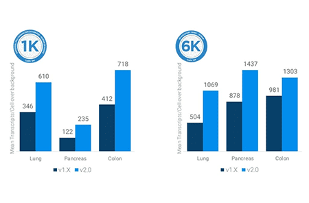
High Sensitivity
Uncover discoveries otherwise missed
Unmatched detection efficiency to deliver the highest number of transcripts and unique genes per cell
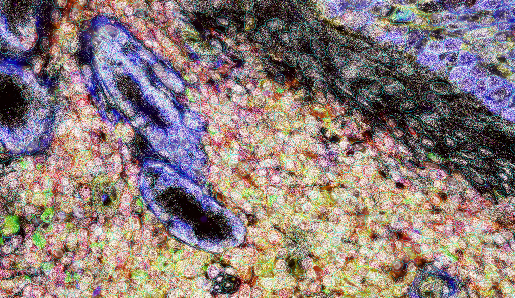
Subcellular Whole Transcriptome Imaging
From inferring interactions to direct biology measurements
First spatial platform to image the entire protein-encoding transcriptome inside every single cell
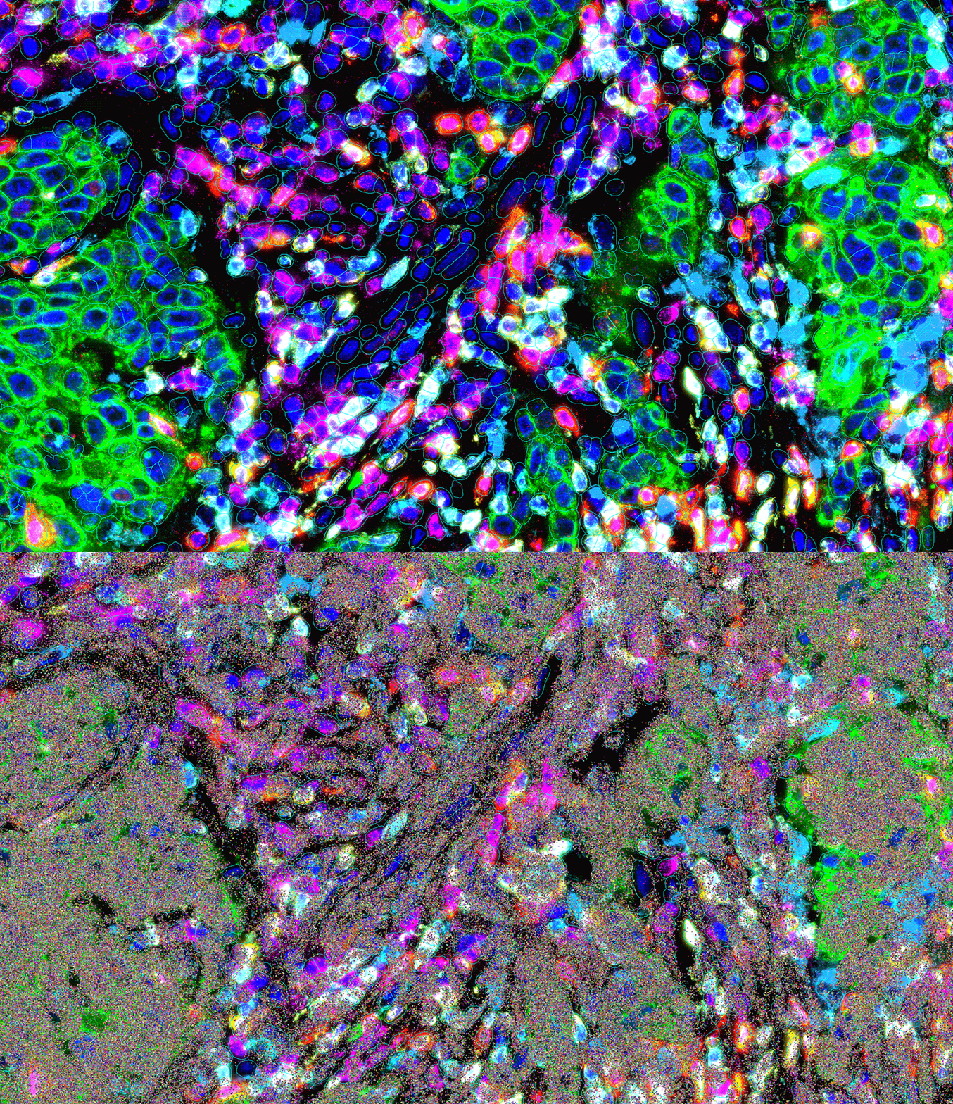
Same-Cell Multiomics
Unlock deeper understanding of cellular function with spatial context
Add another dimension to your data with comprehensive, same-cell RNA and Protein insights
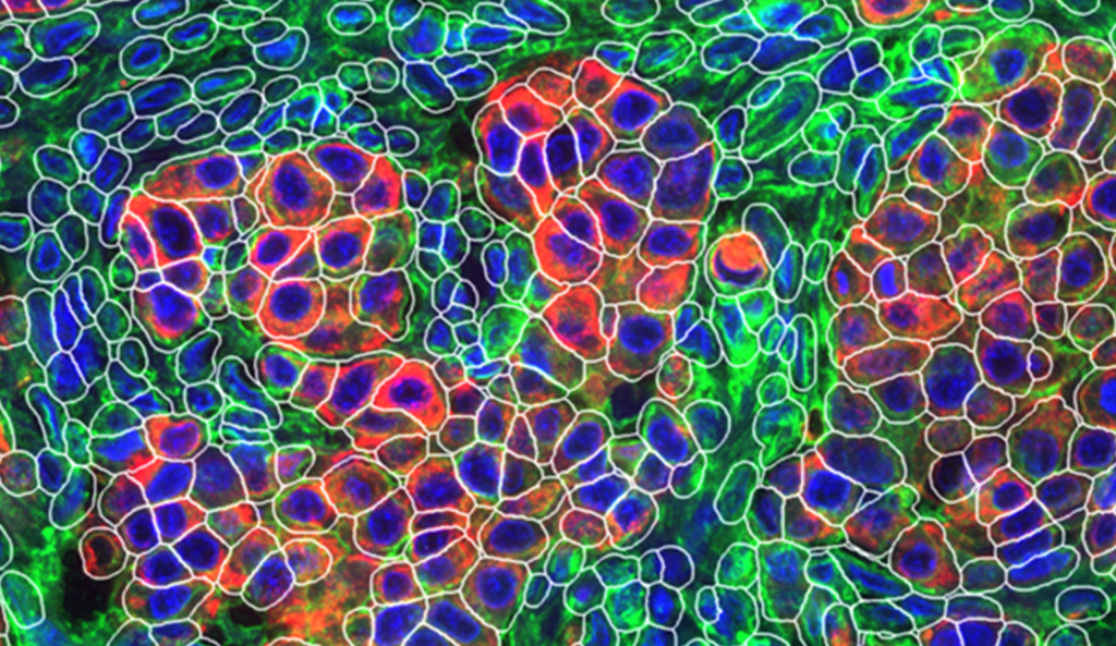
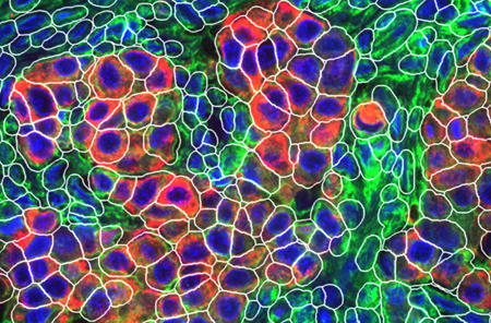
Best 3D Cell Segmentation
Accurately defined boundaries for most reliable biology
Best in class AI algorithms for the most accurate definition of cell boundaries and assignment of transcripts to individual cells
Subcellular Analysis
Make disruptive discoveries for complete layers of biology
Mapping the intracellular landscape with RNA and Protein together at subcellular resolution
Integrated Spatial Informatics
Transform spatial biology into biological insights
The AtoMx Spatial Informatics Platform is the first comprehensive cloud platform, fully integrated with CosMx, for a complete informatics solution from data upload to exploration to export.
Powered by an integrated platform
CosMx SMI combines high‑plex imaging, flexible RNA panels (targeted to whole transcriptome, human and mouse), and AtoMx software for end‑to‑end spatial analysis.

Instrument
Highest sensitivity and plex multiomic instrument accomplished at single-cell and subcellular resolution

Panels & Assays
CosMx RNA and protein panels/assays enable targeted and whole-transcriptome scale spatial discovery.

AtoMx SIP
A unified cloud-native workflow designed to accelerate every stage of spatial data analysis journey.
Trusted by leading researchers in spatial biology
“We are impressed with ease of use with which the CosMx SMI enabled single cell spatial transcriptomics across various tissue types, even in archival FFPE tissue. The high-plex protein assay makes the CosMx SMI a true multiomic solution. The CosMx SMI’s large capture area allows analysis from valuable clinical trial samples using tissue microarrays, enabling characterization of 100s of samples weekly.”

Dr. Nigel B Jamieson
Professor of Surgery, University of Glasgow, UK
“At last count, we have processed 160 samples with the CosMx SMI, including tumor and mouse brain samples. This technology uncovers vital insights into cell biology, enhancing our understanding of both malignant and normal tissues. It is crucial for creating spatial biomarkers and improving patient outcomes. Plus, the equipment and protocols are reliable and user-friendly.”

Dr. Aubrey Thompson
Mayo Clinic Comprehensive Cancer Center, Florida
“The CosMx SMI instrument has become integral to our laboratory’s future direction, and the responsiveness and skill of the customer support for it and the AtoMx SIP has been absolutely fantastic.”

Dr. Grant Kolar
Director, Research Microscopy and Histology Core, St. Louis University School of Medicine
Turning CosMx SMI data into real-world discoveries
Explore spatial transcriptomics workflows designed for translational and discovery research across multiple biological systems and disease areas.

Oncology
Characterize tumor heterogeneity, immune infiltration, and biomarker expression at single-cell resolution directly within intact tissue architecture.

Immunology
Access heterogeneity of immune cell population and disease progression at the cellular and sub-cellular level.

Neuroscience
Investigate neuronal organization, spatial gene expression, and cellular interactions in complex brain tissue samples.

Infectious Disease
Investigate host–pathogen interactions, immune responses, and tissue remodeling with high-plex spatial transcriptomics at single-cell resolution.

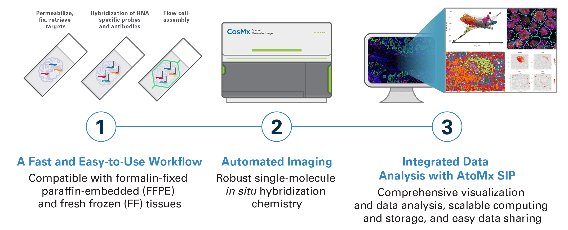
From sample to spatial insights
One spatial molecular imaging platform for high-plex RNA and protein detection, same-cell multiomics, and integrated spatial informatics across FFPE and fresh frozen tissue samples.
Explore the CosMx SMI resource library


Find the right platform for your research
| Platform | 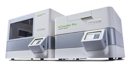 nCounter nCounterLearn more | 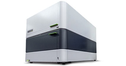 GeoMx GeoMxLearn more | 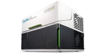 CosMx CosMxLearn more | 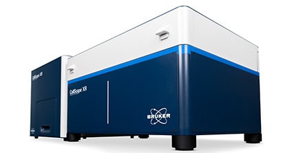 CellScape CellScapeLearn more | 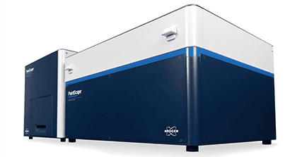 PaintScape PaintScapeLearn more |
| Applications | Rapid and robust targeted RNA and multiomic expression proven for translational and clinical studies | Early biomarker discovery through high-throughput, cost-effective and high-plex RNA and protein screening | High sensitive subcellular imaging of the whole transcriptome for pathway-level spatial insights | Flexible subcellular proteomics for rapid and quantitative spatial phenotyping | High precision, multiplexed direct visualization of the 3D genome |
| Analyte | miRNA, RNA, protein | RNA, protein | RNA, protein | Protein, RNA | DNA |
| Plex level | 800+ RNA 500+ protein | Targeted RNA assays (up to 2000 targets) Whole transcriptome RNA assays Up to 1200 proteins | Target RNA assays (up to 6000 targets) Whole transcriptome RNA assays Up to 64 proteins | Targeted protein assays (up to 34 targets) Customizable assays up to 20 | Targetd DNA Assays (419+ targets) |
| Resolution | Bulk | Multicellular (RNA) Single-cell (protein) | Single-cell Subcellular | Single-cell Subcellular | Single-nuclei |
| Sample compatibility | FFPE, FF, lysate | FFPE, FF | FFPE, FF | FFPE, FF, cell suspension, lysate | Human cell lines, FF tissue (coming soon) |
| Data readout | nCounter (Direct counting) | NGS, nCounter (Direct counting) | Imaging | Imaging | Imaging |
Ready to see what CosMx SMI can do for your research?
Speak with a spatial biology specialist, request a quote, or explore open spatial biology datasets to accelerate your next discovery.
CosMx SMI Frequently Asked Questions
What is CosMx SMI?
CosMx SMI is a high-fidelity spatial molecular imaging platform that enables RNA and protein detection at single-cell and subcellular resolution in FFPE and fresh frozen tissue samples.
What sample types are compatible with CosMx SMI?
CosMx SMI supports formalin-fixed paraffin-embedded (FFPE), fresh frozen tissue samples, organoids and cultured cells enabling flexibility across translational and discovery research workflows.
What RNA assays are available for CosMx SMI?
CosMx Spatial Molecular Imager supports targeted RNA panels up to whole transcriptome workflows, enabling researchers to analyze up to 19,000 RNA targets with spatial context preserved at single-cell and subcellular resolution.
Does CosMx SMI support spatial multiomics?
Yes. CosMx SMI supports same-cell spatial multiomics workflows that enable simultaneous RNA and protein analysis within the same tissue section.
What applications can CosMx SMI support?
CosMx SMI is widely used across oncology, immunology, neuroscience, biomarker discovery, and translational spatial biology research.
What insights can researchers gain from CosMx FFPE datasets?
Researchers can explore cellular heterogeneity, tissue architecture, biomarker expression, immune organization, and pathway-level biology directly within preserved tissue sections.
Does CosMx Spatial Molecular Imager offer grant support for researchers?
Yes. Bruker Spatial Biology provides grant support resources to help researchers incorporate CosMx SMI into funding proposals and spatial biology research applications.
What are the recommended best practices for achieving optimal CosMx® SMI data quality?
CosMx Spatial Molecular Imager best practices include optimizing tissue handling, sample preparation, RNA quality assessment, and workflow execution to maximize sensitivity, spatial resolution, and overall data quality across FFPE and fresh frozen samples.