
Spatial Biology Just Got Bigger
Introducing CellScape™ Whole-Slide Imaging Chamber
Now you can convert any standard microscope slide into a microfluidic chamber for automated multiplex staining, high-resolution imaging, and safe sample archiving. Read more about the expansion of our Precise Spatial Multiplexing capabilities.
Whole-slide spatial biology
Spatial biology is a rapidly growing field that enables exploration and visualization of tissues for a wide variety of applications, including basic research, drug discovery, diagnostic development, and treatment monitoring. Imaging large tissue sections or several samples on the same slide, such as tissue microarrays (TMAs), with whole-slide spatial techniques offers numerous benefits:
- More comprehensive analyses- imaging large areas increases the likelihood of capturing key biological processes.
- Expanded throughput- imaging multiple samples on the same slide enables broader data acquisition at reduced reagent costs.
- Improved statistical power- placing multiple samples on a single slide increases sample numbers, supporting more statistically significant conclusions.
- Enhanced value for clinical applications- imaging larger samples increases the chance of detecting clinically-relevant anomalies in tissues.
- Tissue architectural analysis- whole-slide imaging enables more complex analyses of biological processes in relation to larger tissue structures.
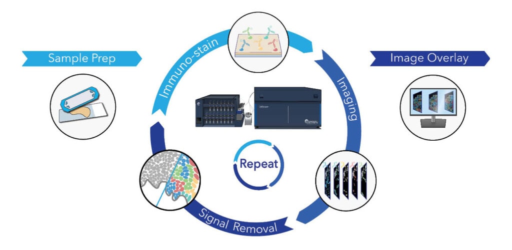
CellScape™ Precise Spatial Multiplexing for highly multiplexed biomarker detection
The CellScape platform enables automated cyclic multiplex immunofluorescence imaging with unprecedented optical resolution needed for quantitative spatial phenotyping. The most versatile instrument for spatial biology, the CellScape utilizes standard fluorescent antibodies for biomarker detection and is compatible with any species or sample type. Leveraging both high-resolution optics and high dynamic range (HDR) imaging, CellScape can detect biomarkers across a wide range of expression levels.
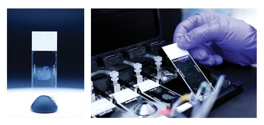
CellScape Whole-Slide Imaging Chamber enables compatibility of CellScape with slides
The CellScape Whole-Slide Imaging Chamber is compatible with any standard histology microscope slide (25 mm by 75 mm). The chamber is composed of a glass coverslip with a removable adhesive on one side. Features of an assembled Slide with Imaging Chamber include:
- 710 mm2 available imaging area
- 50 µL imaging chamber volume
- Automated reagent delivery through imaging chamber inlet and outlet
- Uniform reagent delivery via integrated flow diverters
- Long-term biobanking capability
- Unique sample identification via QR and human readable codes
- Archived FFPE sample compatibility across any species
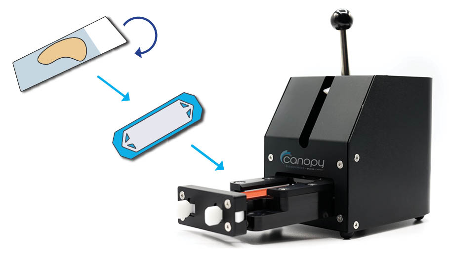
Preparation of a Whole-Slide Imaging Chamber
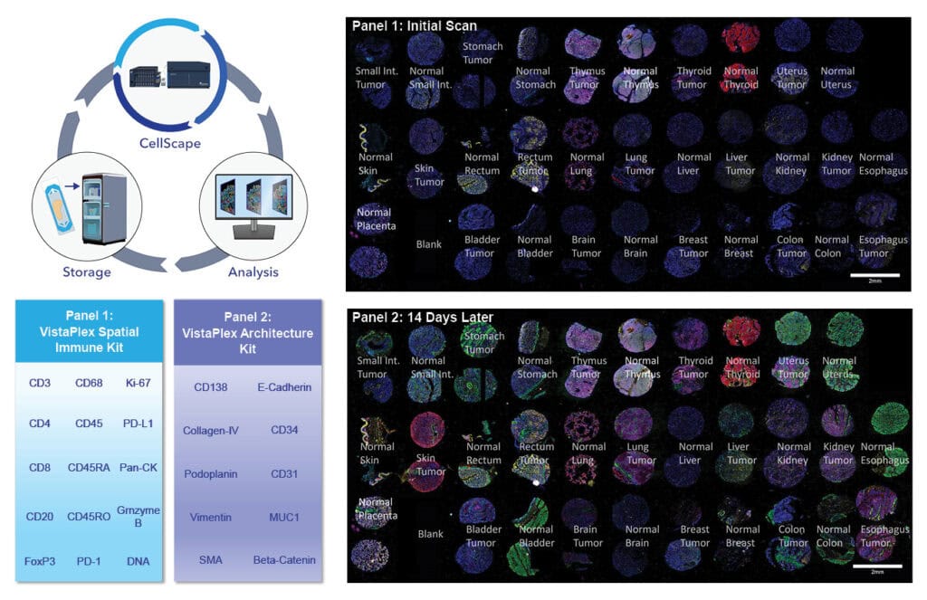
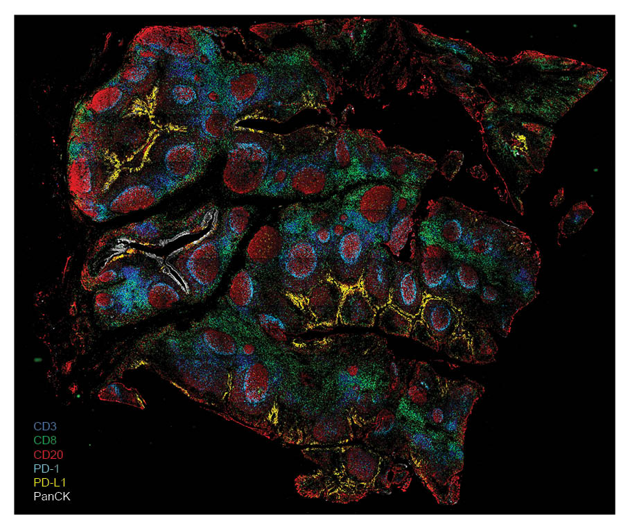
Data-driven assay expansion is particularly useful in fields like cancer biology and immuno-oncology, where it’s important to localize and monitor protein biomarkers to identify diseased tissue, assess immune responses, and monitor treatment progression. In a recent application note, we demonstrate the benefits of data-driven assay expansion to initially map cell populations before expanding the assay with additional staining and imaging to identify specific tissue structures.
We began with human FFPE lung adenocarcinoma samples and used the VistaPlex Spatial Immune Profiling Assay Kit to identify and locate foundational immune and epithelial cell populations.
Cost reduction using CellScape Whole-Slide Imaging Chamber
In addition to providing the maximal viewing area, the CellScape Whole-Slide Imaging Chamber saves costs on reagents compared to other technologies. The platform is compatible with standard, off-the-shelf fluorescent antibodies that are utilized at dilute concentrations, so there is no requirement for custom antibody conjugations or reagent modifications. Additionally, the Whole-Slide Imaging Chamber uses small volumes of reagents, providing an advancement to the original ChipCytometry™ technology utilized by the CellScape platform. The opportunity to combine multiple samples on a single slide further decreases the cost per sample while increasing throughput.
Biobanking and sample reinterrogation using CellScape Whole-Slide Imaging Chambers
A unique feature enabled by CellScape is that samples prepared in Whole-Slide Imaging Chambers are capable of storage and reinterrogation—follow-up staining and imaging of an already-analyzed sample. Reinterrogation provides a flexible and data-driven approach to high-plex spatial biology. Benefits of reinterrogation include:
- On-the-go assay development
- Online quality control of tissue specimens and evaluation of samples for assay suitability
- Ad-hoc assay expansion
- Progressive troubleshooting
- Conservation of precious or rare samples
Below, reinterrogation with a CellScape Whole-Slide Imaging Chamber was demonstrated using two VistaPlex™ Assay Kits, modular and efficient panels for detecting common biomarkers using a CellScape instrument.
Learn more about data-driven assay expansion.
Achieve best-in-class spatial biology data with CellScape Whole-Slide Imaging
The CellScape platform enables high-plex spatial omics with quantitative readouts and flexible reagent choices. Now, the improved sample preparation workflow expands the platform’s capabilities even further by supporting spatial biology without limits, unlocking whole-slide exploration and powerful sample reinterrogation.
Ready to get started? Contact us today for more information or a quote for your new CellScape.


