
Bruker Spatial Biology Blog
Immunology nCounter Oncology
TIS GEP: Use Cases in Sample Stratification, Assessment of Treatment Effect, and as a Component of a Multivariate Biomarker
Introduction to the TIS Gene Expression Profile Innovations in multiplex gene expression analysis are revolutionizing translational research and clinical practices by enabling identification of gene signatures involved in various biological…
Multiomics Spatial Analysis Spatial Biology Spatial Transcriptomics
GeoMx® DSP Spatial Proteogenomics datasets now available on Illumina® BaseSpace™ Sequence Hub
NanoString and Illumina have collaborated to release two new demo datasets of GeoMx Digital Spatial Profiler (DSP) data on the Illumina BaseSpace Sequence Hub (BSSH) cloud. Spatial proteogenomics enables new…

Oncology
Utility of PAM50 GEP Signature as a Biomarker for Exploratory Endpoints in Drug Discovery and Development
The Need for Gene Expression Profiling Gene expression profiling has emerged as a genomic tool that can help decipher the complexity of cancer subtyping. Conceptually, cancer is a fairly simple…
Canopy Biosciences
Why You Need to Increase the Throughput of Your Single-Cell Imaging Experiments
Is your world filled with terms like spatial biology, single-cell analysis, and multiplexed imaging? With a variety of single-cell imaging techniques on the market, it can be time consuming to…
Canopy Biosciences
Developing new protocols to expand the utility of ChipCytometry™
Researcher Spotlight Developing new protocols to expand the utility of ChipCytometry Featuring Sebastian Jarosch, MScInstitute for Medical Microbiology, Immunology, and Hygiene, Technical University of Munich "We have developed a series…
Canopy Biosciences
Profiling the Tumor Microenvironment of Neuroblastoma Using ChipCytometry™
Researcher Spotlight Profiling the Tumor Microenvironment of Neuroblastoma Using ChipCytometry™ Featuring Margarida Neves, MSc, PhD Student, Department of Pathology, UCL Cancer Institute, University College London Department of Translational Medicine, Autolus…

Spatial Biology
Why is the human proteome important?
The Human Protein Atlas is one of the world’s largest biological databases, containing spatial expression profiles for all known proteins expressed in human organs and tissues. It contains a vast…
Spatial Biology
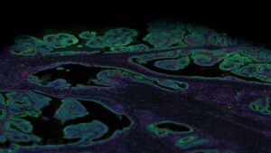
What are immunofluorescence techniques?
Tissue and cell samples constitute the main source of samples for protein or RNA expression analysis in biology and help us understand how cells interact, organize themselves, and drive various…
Spatial Transcriptomics
What is transcriptomics?
The transcriptome is the sum of all the RNA in an organism’s cells and tissues being actively transcribed at any given time from genomic DNA. It includes protein-coding messenger RNA (mRNA)…
Digital Spatial Profiling
What is the principle of immunofluorescence?
Immunofluorescence staining was first used in 1942 and has since remained a highly reliable and powerful technique for a wide range of research and diagnostics. The principle of immunofluorescence is…
CosMx SMI GeoMx DSP
What does nTPM mean in the protein atlas?
The goal of the Human Protein Atlas (HPA) project is to map all human proteins in cells, tissues, and organs. Further, it aims to annotate the function and spatial distribution of…
Digital Spatial Profiling GeoMx DSP
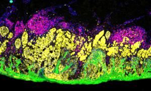
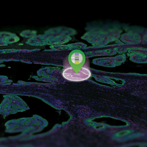
What is a morphology marker?
Understanding tissue morphology is crucial to identify and separate regions of tissue into meaningful biological groupings for image analysis of tissue sections. This is done using morphological markers, most of…